Home
/ White Blood Cells Under Microscope, Frog Blood Cells Light Micrograph Stock Image P243 0008 Science Photo Library : However, their most important difference is that white blood cells are the only ones having a nucleus.
White Blood Cells Under Microscope, Frog Blood Cells Light Micrograph Stock Image P243 0008 Science Photo Library : However, their most important difference is that white blood cells are the only ones having a nucleus.
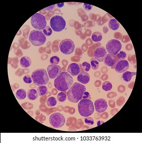
White Blood Cells Under Microscope, Frog Blood Cells Light Micrograph Stock Image P243 0008 Science Photo Library : However, their most important difference is that white blood cells are the only ones having a nucleus.. Human red and white blood cells (including eosinophil, neutrophil) and platelets. Different morphologies can be observed among these cells. Microscopic footage of different white blood cell types found in blood. Therefore, an automatic identification of wbc from. That is the way a doctor can distinguish the ones from the others using the electron microscope when examining your blood specimens.
Reticulocytes are immature red blood cells that are released from the bone marrow. Distinguish the different classes of white blood cells and the conditions under which each would be expected to dominate Under the microscope the most obvious difference is that the white blood cells have an apparent nucleus while the red one have no nucleus actually the somehow large nucleus is from the properties of the white cells. Red blood cells have a central concavity that appears pale under the light microscope. Some of these protect us ag.
White Blood Cells Under Microscope Stock Photos Images Photography Shutterstock from image.shutterstock.com These are the bacteria that cause inflammation and gum disease. Peripheral blood smear results, types of blood cells.white blood cell identification practice:leukocytes: Granulocytes (includes neutrophils, eosinophils and basophils) agranulocytes (includes lymphocytes and monocytes). White blood cells are often characterized as granulocytes or agranulocytes. Several white blood cells from the imaged regions by a 20x conventional microscope (above) and fpm (below). These are divided into two main classes. This classification depends on whether granules can be. The most represented cells in the blood are red blood cells and white blood cells.
In white blood cells, the stain penetrates into the cells and preferentially stainsnuclear material, with weaker staining of the cytoplasm.
A typical blood image usually shows four components: When the monocyte moves into tissue and becomes a macrophage, it becomes even larger with more cytoplasmic granules. White blood cells are the cells of the immune system under the microscope in laboratories. Granulocytes (includes neutrophils, eosinophils and basophils) agranulocytes (includes lymphocytes and monocytes). However, the test can be falsely positive under certain circumstances. Large lymphocytes, monocytes and eosinophils stain a palerblue. Start studying white blood cells under microscope. Stained with monoclonal antibodies for different molecules. Under the microscope the most obvious difference is that the white blood cells have an apparent nucleus while the red one have no nucleus actually the somehow large nucleus is from the properties of the white cells. Distinguish the different classes of white blood cells and the conditions under which each would be expected to dominate Erythrocytes have a lifespan of 120 days. Some of these protect us against bacteria virus and fungus and. Given that all white blood cells are over 5 micrometers in diameter, they are large enough to be seen using a typical optical microscope (compound microscope).
However, their most important difference is that white blood cells are the only ones having a nucleus. Granulocytes (includes neutrophils, eosinophils and basophils) agranulocytes (includes lymphocytes and monocytes). Microscopic footage of different white blood cell types found in blood. If your white blood cell count is truly lower than it should be, then further investigation is warranted to uncover the root cause. These are the bacteria that cause inflammation and gum disease.
White Blood Cell In Blood Smear Analyze By Microscope Stock Photo Picture And Royalty Free Image Image 106735703 from previews.123rf.com Your doctor will monitor your white blood cell count if there is evidence of infection or if you are on medication that may lower your white blood cell count. Peripheral blood smear results, types of blood cells.white blood cell identification practice:leukocytes: Neutrophils, small lymphocytes andplatelets stain most intensely. Red blood cells have a central concavity that appears pale under the light microscope. Hemangioma angioma tumor under microscope. White blood cells are usually bigger than red blood cells and platelets. At 1000x magnification you will be able to see 0.180mm, or 180 microns. This classification depends on whether granules can be.
Microscopic footage of different white blood cell types found in blood.
Stained with monoclonal antibodies for different molecules. Monocytes are the largest type of white blood cell. There is usually only about one white blood cell for every 1,000 red blood cells. Under the microscope the most obvious difference is that the white blood cells have an apparent nucleus while the red one have no nucleus actually the somehow large nucleus is from the properties of the white cells. Human red and white blood cells (including eosinophil, neutrophil) and platelets. This video shows how bacteria and spirochetes attack white blood cells in the mouth. Reticulocytes are immature red blood cells that are released from the bone marrow. Correlate the light and electron microscope images of red and white blood cells; Therefore, an automatic identification of wbc from. A chronically low white blood cell count (leukopenia) can make you vulnerable to bacterial. Your blood is a soup of different kinds of cells, and around 1 % of these cells are immune cells also known as white blood cells. Choose from white blood cell under microscope pics stock illustrations from istock. The type of blood cells to be analyzed.
Quink® appears to make red blood cells (erythrocytes) more visible by weakly binding to theouter surface. The type of blood cells to be analyzed. This classification depends on whether granules can be. White blood cells are usually bigger than red blood cells and platelets. However, the test can be falsely positive under certain circumstances.
White Blood Cells Under Microscope Stock Photos Images Photography Shutterstock from image.shutterstock.com Correspondingly, what can you see at 1000x magnification? The test will become positive if there are a lot of white blood cells in the urine. Start studying white blood cells under microscope. White blood cells are slightly larger, but are much harder to see and require a cell stain or oblique illumination (achieved by adjusting the angle of the light beneath the slide). Our work focuses on the analysis of white blood cells or wbcs. Correlate the light and electron microscope images of red and white blood cells; A chronically low white blood cell count (leukopenia) can make you vulnerable to bacterial. Study of white blood cells are the cells of the immune system under.
As part of your immune system, white blood cells fight disease and are important for the body's defense against infections.
In white blood cells, the stain penetrates into the cells and preferentially stainsnuclear material, with weaker staining of the cytoplasm. Correspondingly, what can you see at 1000x magnification? Quink® appears to make red blood cells (erythrocytes) more visible by weakly binding to theouter surface. Staining with leishman's stain makes it possible to not only easily identify different types of leukocytes, but also count them. Your doctor will monitor your white blood cell count if there is evidence of infection or if you are on medication that may lower your white blood cell count. Study of white blood cells are the cells of the immune system under. The green and red colors come from the staining of the cells with monoclonal. A major distinguishing feature is the presence of granules; Find white blood cell under microscope stock video, 4k footage, and other hd footage from istock. That is the way a doctor can distinguish the ones from the others using the electron microscope when examining your blood specimens. The test will become positive if there are a lot of white blood cells in the urine. Your blood is a soup of different kinds of cells, and around 1 % of these cells are immune cells also known as white blood cells. Red blood cells have a central concavity that appears pale under the light microscope.
